Fascia is not packaging. It is the most densely innervated soft tissue you own — a sensory organ before it is a structural one — and it carries load sideways: a meaningful fraction of a muscle’s force never reaches its own tendon, exiting laterally through the connective sheet instead. The popular claim, that you can manually remodel it with a ball or a roller, is the one part of the story that does not survive contact with the numbers.
What follows is in two halves. Theory is how the tissue works, and what older traditions saw in it long before anyone could film it. Practice is what actually changes it — and what is oversold.
Theory
What the tissue is
Collagen (mostly type I, some III) and elastin fibres suspended in ground substance — water held by proteoglycans and hyaluronan — maintained by fibroblasts and, in the gliding layers, by a specialised cell Carla Stecco’s group named the fasciacyte, whose job is producing hyaluronan.1
Three architectures do real work in everything that follows:
- Dense regular — fibres parallel, built to take load along one line: tendons, aponeuroses, the plantar fascia.
- Dense irregular — the deep fascial sheets, like the fascia lata and the thoracolumbar fascia: sub-layers of parallel collagen stacked at roughly 75–80° to each other,2 so the sheet resists load arriving from several directions at once. This is why single-plane training under-loads it.
- Loose areolar — the wet, slippery layer between the dense ones, where sliding happens and where most of the interesting clinical action lives.
The continuity claim needs stating precisely, because people overreach on it. There is no anatomical seam between the sheet wrapping a muscle, the septum diving between compartments, the periosteum on the bone, and the joint capsule. Those are one continuous connective-tissue body, and the boundaries are created by the dissector’s knife and the textbook’s need for chapters. That much is true. It does not follow that pulling here reliably does something there.
A sensory organ first
The thoracolumbar fascia contains more nerve endings than the muscle underneath it.3 The population is mixed, and each type explains a different sensation. Ruffini endings are slow-adapting and tuned to shear and lateral stretch; their input is associated with reduced sympathetic tone, which is the plausible mechanism for slow, broad work feeling calming rather than merely pleasant. Pacinian corpuscles are fast-adapting and answer vibration and quick change. Golgi receptors sit in the dense, tendinous regions and report tension. And the large majority are thin free nerve endings — polymodal, answering both mechanical load and chemistry, serving both nociception and interoception.
The consequential part is where that traffic goes. A large share of the interoceptive fibres project toward the insula rather than to primary somatosensory cortex,4 so fascial input does not arrive as touch at a coordinate. It arrives as a bodily feeling — warmth, effort, threat, relief, something is off. That routing is why fascial sensation is emotionally coloured and hard to localise, and it is the single most useful fact in the subject.
It is also why stretching over a deep bruise feels electric. Blood outside its vessels is chemically irritating; inflammation drops the mechanical threshold of those endings and recruits silent nociceptors — afferents that are mechanically deaf until inflamed.5 The stretch is not merely louder, it is carried partly by fibres that had no voice before. For a few days, the gain is turned up on anatomy you always had.
Force goes sideways
The textbook model is that a muscle pulls on its tendon and the tendon pulls on the bone. Peter Huijing’s work on epimuscular force transmission showed that a substantial share — on the order of 30% in animal preparations — leaves the muscle laterally, through its wrapping layers, into neighbouring muscles and the compartment wall.6 Force is shared across the sheet before it ever reaches a tendon.
The thoracolumbar fascia is the clearest hub: the latissimus dorsi on one side is continuous, through the sheet, with the opposite gluteus maximus — the posterior oblique sling.7 Pull hard with the right arm and load genuinely arrives at the left hip, through a membrane rather than a muscle. This is the anatomically well-supported end of the fascial-continuity idea.
The other half of load-bearing is elastic storage. The Achilles and plantar fascia stretch on landing and give the energy back on push-off; roughly half the energy of a running stride is recycled this way rather than produced fresh by muscle — the catapult mechanism, extreme in kangaroos and real in humans.8 Collagenous tissue tunes its stiffness to the rate of loading, which is why bouncy, low-amplitude, fast work develops it and slow grinding does not.
The one-way component
Of the two fibres, only one is a renewable resource. Collagen turns over — slowly, but it turns over, and that is what loaded training has to work with. Elastin does not. The gene runs from the third trimester to roughly adolescence and then effectively shuts off,9 and carbon-14 dating of tissue puts the protein’s half-life around 74 years.10 The elastic fibres you have are the ones you built as a child, and the body maintains them the way it maintains nothing else — by not touching them.
The reason is assembly, not the protein. Tropoelastin, the soluble precursor, has to be cross-linked onto a fibrillin scaffold, and that production line only runs during development. Adults still express traces of tropoelastin after injury, but without the line it assembles into disorganised clumps. Wound repair therefore defaults to collagen — which is why scar is strong and does not recoil.
Decay is four attackers on a fixed stock. Mechanical fatigue: pure cycle count, unavoidable — the aorta alone takes on the order of three billion stretch cycles in a lifetime.11 Elastases: elastin-cutting enzymes from immune cells, surging with chronic inflammation and smoking. Ultraviolet light, for the superficial layers, degrading fibres into non-functional clumps. And glycation: sugar welding stiffening cross-links onto the fibres — a protein that is never replaced has a lifetime to accumulate them.
For fascia specifically, distribution matters. Superficial fascia is genuinely elastin-rich — markedly more elastic fibres than the deep sheets12 — which is what lets skin and the subcutaneous layer stretch and snap back. The dense sheets are not: their recoil comes from collagen crimp geometry and the catapult mechanism above, not elastin content. The springiness you can build is collagen architecture and loading rate; the elastin fraction is endowment.
One honest edge: irreplaceable is a today statement, not a law. Recombinant tropoelastin injected into burn and surgical scars produces genuinely new elastin fibres,13 and recent gel and mRNA work shows fibroblasts of any age will build fibres if handed the precursor. All of it is skin-local and early-stage; nothing yet for the deep sheets, arteries, or lungs.
The gliding layer
Between the dense sheets sits loose tissue lubricated by hyaluronan. Stecco’s model: with immobility, overuse, or inflammation, hyaluronan chains aggregate, viscosity rises, and adjacent planes stop sliding freely on each other.1 That is densification — a reversible change in the fluid — and it is a different thing from fibrosis, which is a structural change in the collagen and is not reversible by moving around.
Hyaluronan is non-Newtonian: viscosity falls with shear and with heat. That gives a coherent mechanism for why warmth plus slow cross-fibre work restores glide, and equally for why the effect is temporary — the fluid re-thickens unless you then move through range and keep it sheared. The window is the point, not the technique.
It can contract, slightly
Fascia contains myofibroblasts, and Robert Schleip’s group measured slow autonomous contraction in fascial strips over tens of minutes.14 The forces are far too small and slow to move a joint, but large enough to change tissue stiffness. So the folk claim that tension is held in the tissue has a real substrate — it is just slow, small, and about stiffness rather than posture, which is much less than the claim usually asks for.
The organ nobody could see
In 2018, Neil Theise and colleagues published the first description of the interstitium as a body-wide structure: a network of fluid-filled, collagen-bounded channels running through and around organs, found when a new kind of endoscope — one that images living tissue microscopically, in place — was pointed at a bile duct during routine surgery.15 A century of histology had missed it because fixing tissue to a slide drains the fluid and collapses the channels. The “scientists discover a new organ” headlines wrote themselves.
Radiolab’s episode The Interstitium tells the story properly,16 and its real subject is the blind spot: how millions of scientists and doctors failed to see what was right in front of — and inside — their noses, because the standard way of looking destroyed the thing being looked at. The episode follows two live threads: the channel network as a route by which some cancers spread, and an experiment in which acupuncture needling appeared to activate telocytes, cells that live in these channels. And it lands on the point this piece keeps circling: the “new” discovery sits exactly where Daoist and Chinese medical traditions had been drawing functional maps for two thousand years. Theise frames it as a cultural bridge between ancient and modern medicine — not vindication of any specific claim, but Western anatomy finally seeing a structure another tradition had built theory around for millennia.
Older maps of the same tissue
No tradition has a word for elastin or a diagram of the interstitium, but several kept detailed maps of what this tissue feels like and does, and the overlap with fascial anatomy is close enough to be worth stating carefully.
Chinese medicine’s clearest case is the jingjin — the sinew channels: twelve broad longitudinal bands of muscle, tendon, and connective tissue that parallel the primary meridians and knot at the major joints.17 That is a description of myofascial continuity written some two millennia before the word fascia meant anything. The meridian–fascia correspondence also has a modern research thread:18 Langevin and Yandow’s 2002 mapping put over 80% of arm acupuncture points on intermuscular or intramuscular connective-tissue planes.19 Read that the way this piece reads all continuity maps — a suggestive correspondence between an old functional map and real anatomy, not evidence that qi is a substance. The honest version is that a tradition that spent centuries palpating bodies and drawing lines landed its lines on the planes where the sliding and the sensing actually happen, because that is where fingers find things.
The Daoist reading is more direct and needs no mechanism at all. Daodejing 76: the living are soft and yielding, the dead stiff and hard; the supple belong to life, the rigid to death.20 As phenomenology, that is elastin observed from the outside — infancy as peak compliance because the elastic fibres are new, old age as the one-way stiffening of glycated, fatigued tissue. What the traditions got right is precisely the one-way arrow the biology above establishes; what they could not know is which fibre carries it.
Watch it move
Gil Hedley’s famous fuzz speech belongs beside these — embedding is disabled, but it is worth the click: a dissection-table demonstration of what nightly stillness deposits between sliding surfaces, and why movement is the solvent.
Practice
Load it in every direction
The deep sheets are multi-directional by construction — collagen sub-layers stacked at 75–80° to each other — so rotation, diagonals, and asymmetric loading reach fibre populations that sagittal-plane work never touches. A training week that is all forward-and-back is loading a fraction of the architecture.
Train the spring
The tendon–fascia system adapts to the rate of loading, not just the magnitude. Elastic, low-amplitude, fast work — skipping, low hops, springy ground contact — does more for the Achilles–plantar system than slow calf work. Collagen turnover after a loading bout runs net-negative before it runs net-positive over roughly 36–72 hours,21 so frequency beats intensity: moderate springy work every other day, not occasional heroics. Heroics on an untrained tendon is the standard route into tendinopathy.
Keep the glide
Warmth plus slow, broad, cross-fibre pressure drops hyaluronan viscosity and restores sliding between layers. The change is a window, not a remodel — the fluid re-thickens unless you immediately move through the range you just opened and keep it sheared. Manual work opens the window; movement is what banks it. This is the honest mechanism behind most of what foam rolling and massage genuinely do, and it is worth doing on those terms.
Protect the elastin
Nothing on this list adds elastin; everything on it slows the subtraction. Do not smoke — smoking floods tissue with elastases and is most of why smokers’ skin and lungs age fast. Protect skin from ultraviolet light. Keep blood sugar controlled, because glycation cross-links are forever on a protein that is never replaced. Keep blood pressure in range — every cycle on arterial elastin is wear, and smaller pressure swings are smaller cycles; steady cardiovascular fitness helps exactly here. Keep chronic inflammation down. Topical retinoids nudge modest elastic-fibre repair in skin, and that is about the extent of what can be bought today.
Needles and sinew work
What acupuncture has that survives scrutiny: the planes it targets are real — Langevin’s mapping put most classical points on connective-tissue planes — and needle manipulation measurably grasps and winds subcutaneous connective tissue, deforming fibroblasts for centimetres around the needle.22 That is mechanotransduction, a real physical input to a real sensory organ, and the interstitium and telocyte thread is live research. None of that settles the outcomes literature, which should be read with the usual skepticism; but the tissue mechanics are genuinely interesting, and the tradition was palpating the right layer all along.
On the movement side, the sinew-change tradition is a complete practice built on this tissue. The Yijinjing — literally the sinew-change classic23 — and taiji’s cultivated release, song, amount to slow loaded range, multi-directional, springy, done frequently, with deliberate un-gripping of tension the tissue is holding. It looks a great deal like the modern prescriptions above because both were tuned on the same tissue, one by instruments and one by centuries of practice.
What not to buy
- Manual remodeling of dense fascia. The forces needed to deform the iliotibial band or plantar fascia even about 1% sit far outside anything a hand, elbow, ball, or roller delivers.24 Range-of-motion gains from rolling are real, immediate, short-lived, and neural.
- “Release.” What actually changes: nociceptor sensitivity, autonomic tone, stretch tolerance, and hydration and glide in the loose layer. Every one is a genuine effect; none of them is structural. Calling them structural is what makes honest effects sound like a scam.
- Fascial lines as an atlas. Some continuities have decent dissection support — the posterior oblique sling is one. Others do not. Line maps are a heuristic for where to look next, not a mechanism to cite.25
- Stretching for length. Acute range gains are increased stretch tolerance. Long-term change comes from loaded work at long muscle length over months, and the driver is load.
- Knots. A sensation, not a location — inter-rater agreement on palpating trigger points is poor.26
Two experiments and one caution
While a deep bruise lasts, treat it as free calibration. The stretch that suddenly reads clearly is one you can normally barely feel at all — spend the few days learning the texture of that signal, because the goal is to find it later at normal gain. Most people only ever meet their fascial proprioception when it is inflamed, and conclude it is a pain channel.
The second experiment is the glide window. After warmth and slow shear work, note how long the loose, easy quality lasts if you sit down versus if you move through full range for ten minutes. If the model above is right, the difference should be obvious and repeatable.
The caution: for the first 48–72 hours after a deep contusion, gentle pain-free range only. Forceful early work on a fresh intramuscular haematoma is the classic setup for re-bleeding and for myositis ossificans, where the haematoma calcifies into bone inside the muscle.27 Rare, but it punishes precisely the instinct this piece cultivates, because the stretching feels productive at the moment it is least advisable.
References
Reference summary — Anatomy Trains
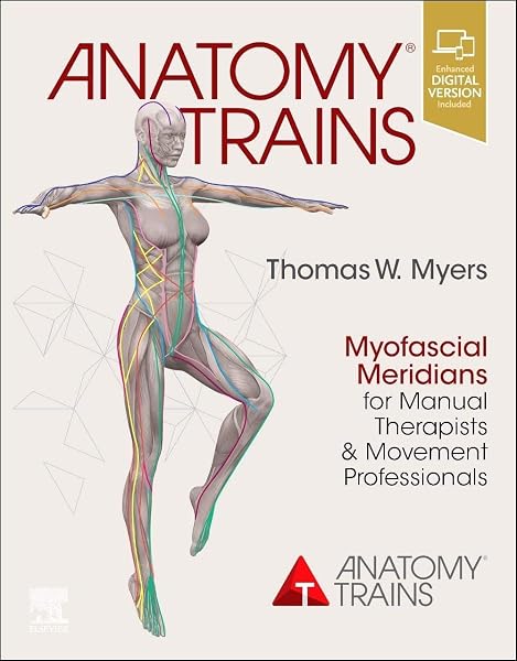
A book that sits under this whole subject gets more than a footnote. Thomas Myers’s Anatomy Trains (buy it · anatomytrains.com) argues that one-muscle-at-a-time anatomy is an artifact of dissection, not of bodies: the textbook cuts fascia away to reveal the muscle, and the stuff being cut away is doing structural work. He organises the continuities into about twelve named lines, and — to his credit — states rules for what counts as one, which makes the map testable rather than mystical.
What holds up: the tissue is continuous and force does cross it, and the book gave clinicians a shared vocabulary for something they kept seeing, which made it discussable and therefore testable. What to distrust: fascia is continuous everywhere, so a dissection looking for a line will always find one — the real question is whether tension meaningfully travels that route, and most of the supporting work does not answer it. The Deep Front Line, the one running tongue → hyoid → diaphragm → psoas that this piece keeps circling, is the deepest and least supported of the lines. Its seductiveness is the warning, not the evidence.
The verdict this piece uses: a map, not a mechanism. A good map — it points attention at continuities conventional anatomy hides. Not a mechanism — it does not establish that tension travels those routes in a living body. Holding both at once is the skill.
Sources
This is a living document.
Further reading, cited nowhere above but worth the time: Jean-Claude Guimberteau, Strolling under the Skin (2005), and the endoscopic archive at endovivo.com; Gil Hedley, the fuzz speech.
-
Stecco et al., The fasciacytes: a new cell devoted to fascial gliding regulation, Clinical Anatomy (2018). DOI 10.1002/ca.23072 ↩ ↩2
-
Benetazzo, Bizzego, De Caro, Frigo, Guidolin & Stecco, 3D reconstruction of the crural and thoracolumbar fasciae, Surgical and Radiologic Anatomy (2011). DOI 10.1007/s00276-010-0757-7
“The angle between the fibers of adjacent layers is about 78°.” ↩
-
Tesarz, Hoheisel, Wiedenhöfer & Mense, Sensory innervation of the thoracolumbar fascia in rats and humans, Neuroscience (2011). DOI 10.1016/j.neuroscience.2011.07.066 ↩
-
Schleip & Jäger, Interoception: a new correlate for intricate connections between fascial receptors, emotion and self recognition, in Fascia: The Tensional Network of the Human Body (2012). Chapter text ↩
-
Mense & Hoheisel, Evidence for the existence of nociceptors in rat thoracolumbar fascia, Journal of Bodywork and Movement Therapies (2016). PubMed 27634088 ↩
-
Huijing, Epimuscular myofascial force transmission: a historical review and implications for new research, Journal of Biomechanics (2009). DOI 10.1016/j.jbiomech.2008.09.027 — the ~30% lateral share is from animal preparations; the human in-vivo analogue is not established. ↩
-
Vleeming, Pool-Goudzwaard, Stoeckart, van Wingerden & Snijders, The posterior layer of the thoracolumbar fascia: its function in load transfer from spine to legs, Spine (1995). PubMed 7701385 ↩
-
Alexander & Vernon, The mechanics of hopping by kangaroos (Macropodidae), Journal of Zoology (1975). DOI 10.1111/j.1469-7998.1975.tb05983.x — the wallaby saving is calculated at about 40%; the “roughly half” figure in the text is the looser popular rounding. ↩
-
Emerging mechanisms of elastin transcriptional regulation, American Journal of Physiology — Cell Physiology (2022). DOI 10.1152/ajpcell.00228.2022
“Elastin gene expression and protein synthesis occur within a narrow timeframe initiating late in embryonic development and terminating in adolescence with essentially no de novo elastin production throughout adult life.” ↩
-
Shapiro et al., Marked longevity of human lung parenchymal elastic fibers deduced from prevalence of D-aspartate and nuclear weapons-related radiocarbon, Journal of Clinical Investigation (1991). DOI 10.1172/JCI115204
“The calculated mean carbon residence time in elastin was 74 years (95% confidence limits, 40–174 years).” Strictly a residence time, not a half-life. ↩
-
Cocciolone et al., Elastin, arterial mechanics, and cardiovascular disease, American Journal of Physiology — Heart and Circulatory Physiology (2018). PMC6139627 ↩
-
Pirri et al., Elastic fibres in the subcutaneous tissue: is there a difference between superficial and muscular fascia? A cadaver study, Skin Research and Technology (2022). DOI 10.1111/srt.13084 ↩
-
Treatment of burn and surgical wounds with recombinant human tropoelastin produces new elastin fibers in scars (2017). PubMed 28221299 ↩
-
Schleip et al., Active contractile properties of fascia, Clinical Anatomy (2019). DOI 10.1002/ca.23391 ↩
-
Benias, Theise et al., Structure and distribution of an unrecognized interstitium in human tissues, Scientific Reports (2018). DOI 10.1038/s41598-018-23062-6 ↩
-
Radiolab, The Interstitium, reported by Lulu Miller and Jenn Brandel. ↩
-
Huangdi Neijing, Lingshu — the jingjin (sinew channels) chapters. Lingshu Jing — cited from secondary sources; primary translation check pending. ↩
-
Bai et al., Review of evidence suggesting that the fascia network could be the anatomical basis for acupoints and meridians in the human body, Evidence-Based Complementary and Alternative Medicine (2011). DOI 10.1155/2011/260510 ↩
-
Langevin & Yandow, Relationship of acupuncture points and meridians to connective tissue planes, The Anatomical Record (2002). DOI 10.1002/ar.10185 ↩
-
Laozi, Daodejing, chapter 76 — the living are soft and yielding; the dead, stiff and hard. ↩
-
Magnusson, Langberg & Kjaer, The pathogenesis of tendinopathy: balancing the response to loading, Nature Reviews Rheumatology (2010). DOI 10.1038/nrrheum.2010.43 — source of the 36–72 hour turnover window. ↩
-
Langevin, Churchill & Cipolla, Mechanical signaling through connective tissue: a mechanism for the therapeutic effect of acupuncture, The FASEB Journal (2001). DOI 10.1096/fj.01-0015hyp ↩
-
The Yijin Jing (sinew-change classic) tradition of conditioning practice. ↩
-
Chaudhry, Schleip, Ji, Bukiet, Maney & Findley, Three-dimensional mathematical model for deformation of human fasciae in manual therapy, Journal of the American Osteopathic Association (2008). DOI 10.7556/jaoa.2008.108.8.379 — source of the ~1% deformation threshold. ↩
-
Thomas Myers, Anatomy Trains — myofascial meridians: the continuous fascial lines that link muscles into functional chains across the body. ↩
-
Lucas, Macaskill, Irwig, Moran & Bogduk, Reliability of physical examination for diagnosis of myofascial trigger points: a systematic review of the literature, The Clinical Journal of Pain (2009). PubMed 19158550
“Physical examination cannot currently be recommended as a reliable test for the diagnosis of trigger points.” ↩
-
Myositis ossificans of the quadriceps, Physiopedia — heterotopic bone formation following contusion and intramuscular haematoma. ↩